
 A SCHOLARLY BOOK REPOSITORY
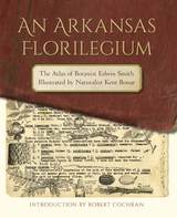
Thirty-five years later, with Smith retired and Bonar long gone from the park service but still drawing, Bonar’s weathered and battered copy of the atlas was seized by a diverse cadre of amateur admirers motivated by fears of its damage or loss. Their fears were certainly justified; after all, the pages were now jammed to the margins with some 3,500 drawings, and the volume had already survived one accidental dunking in an Ozark stream.
An Arkansas Florilegium brings Smith’s and Bonar’s knowledge and lifelong diligence to the world in this unique mix of art, science, and Arkansas saga.

Edited by historian Nancy S. Seasholes, this landmark volume captures all aspects of Boston’s past in a series of fifty-seven stunning full-color spreads. Each section features newly created thematic maps that focus on moments and topics in that history. These maps are accompanied by hundreds of historical and contemporary illustrations and explanatory text from historians and other expert contributors. They illuminate a wide range of topics including Boston’s physical and economic development, changing demography, and social and cultural life. In lavishly produced detail, The Atlas of Boston History offers a vivid, refreshing perspective on the development of this iconic American city.
Contributors
Robert J. Allison, Robert Charles Anderson, John Avault, Joseph Bagley, Charles Bahne, Laurie Baise, J. L. Bell, Rebekah Bryer, Aubrey Butts, Benjamin L. Carp, Amy D. Finstein, Gerald Gamm, Richard Garver, Katherine Grandjean, Michelle Granshaw, James Green, Dean Grodzins, Karl Haglund, Ruth-Ann M. Harris, Arthur Krim, Stephanie Kruel, Kerima M. Lewis, Noam Maggor, Dane A. Morrison, James C. O’Connell, Mark Peterson, Marshall Pontrelli, Gayle Sawtelle, Nancy S. Seasholes, Reed Ueda, Lawrence J. Vale, Jim Vrabel, Sam Bass Warner, Jay Wickersham, and Susan Wilson
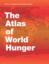
Earlier this year, President Obama declared one of his top priorities to be “making sure that people are able to get enough to eat.” The United States spends about five billion dollars on food aid and related programs each year, but still, both domestically and internationally, millions of people are hungry. In 2006, the Food and Agricultural Organization of the United Nations counted 850 million hungry people worldwide, but as food prices soared, an additional 100 million or more who were vulnerable succumbed to food insecurity.
If hunger were simply a matter of food production, no one would go without. There is more than enough food produced annually to provide every living person with a healthy diet, yet so many suffer from food shortages, unsafe water, and malnutrition every year. That’s because hunger is a complex political, economic, and ecological phenomenon. The interplay of these forces produces a geography of hunger that Thomas J. Bassett and Alex Winter-Nelson illuminate in this empowering book. The Atlas of World Hunger uses a conceptual framework informed by geography and agricultural economics to present a hunger index that combines food availability, household access, and nutritional outcomes into a single tool—one that delivers a fuller understanding of the scope of global hunger, its underlying mechanisms, and the ways in which the goals for ending hunger can be achieved. The first depiction of the geography of hunger worldwide, the Atlas will be an important resource for teachers, students, and anyone else interested in understanding the geography and causes of hunger. This knowledge, the authors argue, is a critical first step toward eliminating unnecessary suffering in a world of plenty.
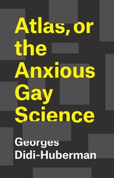
In this illustrated exploration of Warburg and his great work, Georges Didi-Huberman leaps from Mnemosyne Atlas into a set of musings on the relation between suffering and knowledge in Western thought, and on the creative results of associative thinking. Deploying writing that delights in dramatic jump cuts reminiscent of Warburg’s idiosyncratic juxtapositions, and drawing on a set of sources that ranges from ancient Babylon to Walter Benjamin, Atlas, or the Anxious Gay Science is rich in Didi-Huberman’s trademark combination of elan and insight.
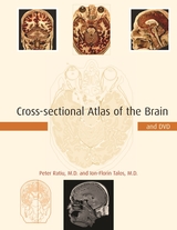
Cross-sectional Atlas of the Brain provides for the first time a set of high-resolution color cross-sections of the human brain (six times higher than that of the only complete data set available to date), each image accompanied by state-of-the-art MRI and CT scans of the same specimen. The sections were made at an interval of 147 micrometers of frozen tissue, virtually artifact free, with the blood vessels filled at sub-millimeter level. The more than two hundred detailed and fully annotated images in this atlas provide a complete body of reference to the gross anatomy of the brain. The accompanying line drawings of these images provide a roadmap for easy orientation.
The unparalleled resolution of the images also made it possible to derive cross-sections of the same specimen in all standard orientations--sagittal, coronal, and axial--through multi-planar computer-aided reformatting. This feature, which eliminates inter-subject variability, has never before been available in an anatomical atlas and makes the atlas especially useful for identifying and following anatomical structures in each plane. About the Companion DVD(View a sample in PDF format)
While the book itself contains 93 images (44 axial, 28 coronal, and 21 sagittal), the DVD contains the complete series of 1,481 axial images from one anatomic specimen from which the 44 axial images in the book were selected. These images were made at a resolution of 1525x1146 or 147 µm/pixel with a digital camera. The axial images are accompanied by 1,528 sagittal and 1,146 coronal images that were made by reformatting and reslicing the axial images. By placing these images side-by-side-by-side the DVD allows the user to see a particular region of the brain in all three orientations-axial, sagittal and coronal-simultaneously. These images are further accompanied by radiologic data. The DVD also allows the user to view a synchronized slide show of the images in all three planes. Images on the DVD that also appear in the book are highlighted with a blue background.
Cross-sectional Atlas of the Brain will be an essential reference for neuroscientists and clinicians (neurologists, radiologists, and neurosurgeons).
Rapidly developing diagnostic and therapeutic methods involving direct contact with the human fetus—fetoscopy, fetal surgery, ultrasonic scanning— demand a precise knowledge of normal structural development during gestation. Toward achieving that goal of precision, Drs. Robert Shapiro and Franklin Robinson have created an atlas described by Richard L. Sidman as “a solid piece of research, executed with considerable esthetic as well as scholarly finesse, and [which] will serve as the definitive study on an important aspect of human fetal development.”
The authors have documented the early development of the human skull in terms of gross size, shape, and the behavior of the individual bones com posing the skull with reference to their ossification centers, ossification rates, and relationships. The data are presented in very high quality photographs and radiographs of the dried skull in several relevant orientations, low magnification color photomicrographs of well sectioned and stained specimens, and color photographs of an unusually fine series of transilluminated skulls prepared by the Spalteholz method. Line drawings are also presented to assist in interpretation.
The atlas is organized according to gestational age, and a tabular summary is given of the 63 specimens ranging in age from ten to forty fetal weeks.
This will be the basic normative standard reference for studies on develop mental skeletal disorders of the head and neck; it will be useful as well in the study of developmental brain diseases. Radiologists engaged in visualizing the fetus and diagnosing fetal diseases in situ by ultrasound, computerized tomography, and other methods will find this an invaluable tool.
The expansive and intricate Atlas Coelestis, created by Johann Doppelmayr in 1742, set out to record everything known about astronomy at the time, covering constellations, planets, moons, comets, and more, all rendered in exquisite detail. Through stunning illustrations, historical notes, and scientific explanations, Phenomena contextualizes Doppelmayr’s atlas and creates a spectacular handbook to the heavens.
Phenomena begins by introducing Doppelmayr’s life and work, placing his extraordinary cosmic atlas in the context of discoveries made in the Renaissance and Enlightenment and highlighting the significance of its publication. This oversized book presents thirty beautifully illustrated and richly annotated plates, covering all the fundamentals of astronomy—from the dimensions of the solar system to the phases of the moon and the courses of comets. Each plate is accompanied by expert analysis from astronomer Giles Sparrow, who deftly presents Doppelmayr’s references and cosmological work to a modern audience. Each plate is carefully deconstructed, isolating key stars, planets, orbits, and moons for in-depth exploration. A conclusion reflects on the development of astronomy since the publication of the Atlas and traces the course of the science up to the present day. Following the conclusion is a timeline of key discoveries from ancient times onward along with short biographies of the key players in this history.
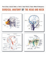
Surgical Anatomy of the Head and Neck was immediately hailed as indispensable when it was first published in 2001. In demand ever since, this classic surgical atlas—packed with more than 700 exceptional drawings, 537 of them in full color, by an internationally noted medical illustrator—is now available again, with an extensive new index, after years of being out of print.
Here is a surgeon’s-eye view of all anatomic details, from the upper thorax to the crown. Ideal for both surgery and test preparation, this volume features special boxed sections that focus on the surgical significance of each anatomical structure. Every illustration is clearly labeled with key anatomic landmarks, and a user-friendly design allows quick reference. This volume is an invaluable resource for surgeons, residents, and medical students.
READERS
Browse our collection.
PUBLISHERS
See BiblioVault's publisher services.
STUDENT SERVICES
Files for college accessibility offices.
RECENTLY PUBLISHED


UChicago Accessibility Resources
home | accessibility | search | about | contact us
BiblioVault ® 2001 - 2024
The University of Chicago Press