
 A SCHOLARLY BOOK REPOSITORY
"It is my hope," writes the author in his preface, "that the material collected in this monograph will provide a new perspective in some areas for those students, biologists, and physicians interested in the kidney but too involved with their special trees to notice some of the changes occurring in the forest in which they toil."
In the first up-to-date compendium to correlate the changes in kidney structure and function from the onset of organogenesis to the end of childhood growth, Dr. Wallace McCrory presents a new aspect of developmental pediatrics, skillfully explains the clinical enigmas surrounding the immature kidney, and suggests possible research areas for productive exploration. Clearly documented with tables and illustrations, the study synthesizes relevant new knowledge from the fields of embryology, biochemistry, and renal and growth physiology as a means of stimulating reappraisals of the current concepts of the pathophysiology of many childhood renal diseases. Included are reproductions of reconstructed micro dissections of the early stages in the developing human kidney taken from Dr. Jean Oliver's monograph Nephrons and Kidneys.
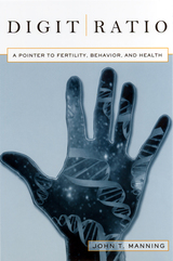
It has been known for more than a century that men and women tend to differ in the relative lengths of their index (2D) and ring (4D) fingers, which upon casual observation seem fairly symmetrical. Men on average have fourth digits longer than their second digits, while women typically have the opposite. Digit ratios are unique in that they are fixed before birth, while other sexually dimorphic variables are fixed after puberty, and the same genes that control for finger length also control the development of the sex organs. The 2D:4D ratio is the only prenatal sexually dimorphic trait that measurably explains conditions linking testosterone, estrogen, and human development; the study of the ratio broadens our view of human ability, talent, behavior, disposition, health, and fertility. In this book, Manning presents evidence for how 2D:4D correlates with traits ranging from sperm counts, family size, musical genius, and sporting prowess, to autism, depression, homosexuality, heart attacks, and breast cancer, traits that are all linked with early exposure to sex hormones.
Rapidly developing diagnostic and therapeutic methods involving direct contact with the human fetus—fetoscopy, fetal surgery, ultrasonic scanning— demand a precise knowledge of normal structural development during gestation. Toward achieving that goal of precision, Drs. Robert Shapiro and Franklin Robinson have created an atlas described by Richard L. Sidman as “a solid piece of research, executed with considerable esthetic as well as scholarly finesse, and [which] will serve as the definitive study on an important aspect of human fetal development.”
The authors have documented the early development of the human skull in terms of gross size, shape, and the behavior of the individual bones com posing the skull with reference to their ossification centers, ossification rates, and relationships. The data are presented in very high quality photographs and radiographs of the dried skull in several relevant orientations, low magnification color photomicrographs of well sectioned and stained specimens, and color photographs of an unusually fine series of transilluminated skulls prepared by the Spalteholz method. Line drawings are also presented to assist in interpretation.
The atlas is organized according to gestational age, and a tabular summary is given of the 63 specimens ranging in age from ten to forty fetal weeks.
This will be the basic normative standard reference for studies on develop mental skeletal disorders of the head and neck; it will be useful as well in the study of developmental brain diseases. Radiologists engaged in visualizing the fetus and diagnosing fetal diseases in situ by ultrasound, computerized tomography, and other methods will find this an invaluable tool.
Too tiny to see with the naked eye, the human embryo was just a hypothesis until the microscope made observation of embryonic development possible. This changed forever our view of the minuscule cluster of cells that looms large in questions about the meaning of life. Embryos under the Microscope examines how our scientific understanding of the embryo has evolved from the earliest speculations of natural philosophers to today’s biological engineering, with its many prospects for life-enhancing therapies. Jane Maienschein shows that research on embryos has always revealed possibilities that appear promising to some but deeply frightening to others, and she makes a persuasive case that public understanding must be informed by up-to-date scientific findings.
Direct observation of embryos greatly expanded knowledge but also led to disagreements over what investigators were seeing. Biologists confirmed that embryos are living organisms undergoing rapid change and are not in any sense functioning persons. They do not feel pain or have any capacity to think until very late stages of fetal development. New information about DNA led to discoveries about embryonic regulation of genetic inheritance, as well as evolutionary relationships among species. Scientists have learned how to manipulate embryos in the lab, taking them apart, reconstructing them, and even synthesizing—practically from scratch—cells, body parts, and maybe someday entire embryos. Showing how we have learned what we now know about the biology of embryos, Maienschein changes our view of what it means to be alive.
READERS
Browse our collection.
PUBLISHERS
See BiblioVault's publisher services.
STUDENT SERVICES
Files for college accessibility offices.
RECENTLY PUBLISHED

struction of Metaphysics")


UChicago Accessibility Resources
home | accessibility | search | about | contact us
BiblioVault ® 2001 - 2024
The University of Chicago Press