The Comparative Anatomy and Histology of the Cerebellum was first published in 1970. Minnesota Archive Editions uses digital technology to make long-unavailable books once again accessible, and are published unaltered from the original University of Minnesota Press editions.
This is the second volume of the late Dr. Larsell's comprehensive monograph on the cerebellum, the first volume of which is described below. A third volume, on the human cerebellum, will be published by the University of Minnesota Press next spring to complete the work.
This second volume deals with the morphogenetic development and morphology of the cerebellum of all orders of mammals from monotremes through apes. The descriptions cover the cerebellum in about forty species with special emphasis on the cerebellum of the albino rate, rabbit, cat, and rhesus monkey. Dr. Larsell's comparative anatomical studies over a period of many years led to the conclusion that fundamentally the mammalian cerebellum is composed of ten subdivisions. With few exceptions (the smallest and most primitive cerebella) the subdivisions are identified in all mammals. The descriptions of the cerebella are based on the author's personal investigations but the relevant literature is thoroughly reviewed also.

The Comparative Anatomy and Histology of the Cerebellum was first published in 1972. Minnesota Archive Editions uses digital technology to make long-unavailable books once again accessible, and are published unaltered from the original University of Minnesota Press editions.
This is the third and final volume of the late Dr. Larsell's definitive work on the cerebellum, brought to completion for publication by Dr. Jansen. Two additional contributing authors for this volume are Enrico Mugnaini, M.D., and Helge K. Korneliussen, M.D.
The first section of this volume deals with the morphology of the human cerebellum. The morphogenetic development, the fissure formation, and the differentiation of the cerebellar lobules are described in detail, and followed by a comprehensive account of the adult cerebellum, its lobes and lobules. It is shown that the ten major lobules which Dr. Larsell distinguished in other mammals are recognizable also in man.
Chapters on the cerebellum connections include detailed accounts of all afferent and efferent cerebellar tracts. A subsequent chapter, by Drs. Jansen and Korneliussen, is devoted to the fundamental plan of cerebellar organization. The final chapters, by Dr. Mugnaini, deal with the histology and cytology of the cerebellar cortex. A comprehensive account is given of electron micrographs, a virtual atlas of the ultrastructure of the cerebellar cortex, illustrate the description.
• 26 chapters covering all basic tissues and major organs/organ systems of human histology
• More than 1100 high quality histological images on 724 pages
• 236 image-based multiple choice review questions
• Volume 1: Introduction, Epithelia, Connective Tissue, Cartilage, Bone, Blood
• Volume 2: Muscle, Nervous System, Eye, Ear, Circulatory System, Lymphatic System
• Volume 3: Respiratory System, Integument, Oral Glands, Oral Cavity, Gastrointestinal Tract, Liver, Gall Bladder & Pancreas
• Volume 4: Endocrine Organs, Urinary System, Male Reproductive System, Female Reproductive System, Mammary Gland
“Don MacCallum’s Michigan Histology” is complemented by the Michigan Histology Virtual Slide Collection, which can be accessed for free via the Internet at http://histology.sites.uofmhosting.net
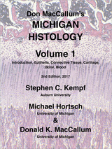
Volume 1 of this 4 Volume lab manual set is concerned with the Histology of human tissues. It covers Epithelia, General Connective Tissue, Cartilage, Bone and Blood with a descriptive text and pictures.
• 26 chapters covering all basic tissues and major organs/organ systems of human histology
• More than 1100 high quality histological images on 724 pages
• 236 image-based multiple choice review questions
• Volume 1: Introduction, Epithelia, Connective Tissue, Cartilage, Bone, Blood
• Volume 2: Muscle, Nervous System, Eye, Ear, Circulatory System, Lymphatic System
• Volume 3: Respiratory System, Integument, Oral Glands, Oral Cavity, Gastrointestinal Tract, Liver, Gall Bladder & Pancreas
• Volume 4: Endocrine Organs, Urinary System, Male Reproductive System, Female Reproductive System, Mammary Gland
“Don MacCallum’s Michigan Histology” is complemented by the Michigan Histology Virtual Slide Collection, which can be accessed for free via the Internet at http://histology.sites.uofmhosting.net
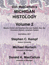
Volume 2 of this 4 Volume lab manual set is concerned with the Histology of human tissues. It covers Muscle, Nervous System, Eye, Ear, Circulatory System, and Lymphatic System with a descriptive text and pictures.
• 26 chapters covering all basic tissues and major organs/organ systems of human histology
• More than 1100 high quality histological images on 724 pages
• 236 image-based multiple choice review questions
• Volume 1: Introduction, Epithelia, Connective Tissue, Cartilage, Bone, Blood
• Volume 2: Muscle, Nervous System, Eye, Ear, Circulatory System, Lymphatic System
• Volume 3: Respiratory System, Integument, Oral Glands, Oral Cavity, Gastrointestinal Tract, Liver, Gall Bladder & Pancreas
• Volume 4: Endocrine Organs, Urinary System, Male Reproductive System, Female Reproductive System, Mammary Gland
“Don MacCallum’s Michigan Histology” is complemented by the Michigan Histology Virtual Slide Collection, which can be accessed for free via the Internet at http://histology.sites.uofmhosting.net
Volume 3 of this 4 Volume lab manual set is concerned with the Histology of human tissues. It covers Respiratory System, Integument, Oral Glands, Oral Cavity, Gastrointestinal Tract, Liver, Gall Bladder, and Pancreas with a descriptive text and pictures.
• 26 chapters covering all basic tissues and major organs/organ systems of human histology
• More than 1100 high quality histological images on 724 pages
• 236 image-based multiple choice review questions
• Volume 1: Introduction, Epithelia, Connective Tissue, Cartilage, Bone, Blood
• Volume 2: Muscle, Nervous System, Eye, Ear, Circulatory System, Lymphatic System
• Volume 3: Respiratory System, Integument, Oral Glands, Oral Cavity, Gastrointestinal Tract, Liver, Gall Bladder & Pancreas
• Volume 4: Endocrine Organs, Urinary System, Male Reproductive System, Female Reproductive System, Mammary Gland
“Don MacCallum’s Michigan Histology” is complemented by the Michigan Histology Virtual Slide Collection, which can be accessed for free via the Internet at http://histology.sites.uofmhosting.net
Volume 4 of this 4 Volume lab manual set is concerned with the Histology of human tissues. It covers Endocrine Organs, Urinary System, Male Reproductive System, Female Reproductive System, and the Mammary Gland with a descriptive text and pictures.
Ever since the threads of seventeenth-century natural philosophy began to coalesce into an understanding of the natural world, printed artifacts such as laboratory notebooks, research journals, college textbooks, and popular paperbacks have been instrumental to the development of what we think of today as “science.” But just as the history of science involves more than recording discoveries, so too does the study of print culture extend beyond the mere cataloguing of books. In both disciplines, researchers attempt to comprehend how social structures of power, reputation, and meaning permeate both the written record and the intellectual scaffolding through which scientific debate takes place.
Science in Print brings together scholars from the fields of print culture, environmental history, science and technology studies, medical history, and library and information studies. This ambitious volume paints a rich picture of those tools and techniques of printing, publishing, and reading that shaped the ideas and practices that grew into modern science, from the days of the Royal Society of London in the late 1600s to the beginning of the modern U.S. environmental movement in the early 1960s.
READERS
Browse our collection.
PUBLISHERS
See BiblioVault's publisher services.
STUDENT SERVICES
Files for college accessibility offices.
UChicago Accessibility Resources
home | accessibility | search | about | contact us
BiblioVault ® 2001 - 2024
The University of Chicago Press


