Results by title
5 books match the query 'Diagnostic Imaging'

CT Suite
The Work of Diagnosis in the Age of Noninvasive CuttingBarry F. Saunders
Duke University Press, 2008
Saunders’s analyses are informed by strands of cultural history and theory including art historical critiques of realist representation, Walter Benjamin’s concerns about violence in “mechanical reproduction,” and tropes of detective fiction such as intrigue, the case, and the culprit. Saunders analyzes the diagnostic “gaze” of medical personnel reading images at the viewbox, the two-dimensional images or slices of the human body rendered by the scanner, methods of archiving images, and the use of scans as pedagogical tools in clinical conferences. Bringing cloistered diagnostic practices into public view, he reveals the customs and the social and professional hierarchies that are formulated and negotiated around the weighty presence of the CT scanner. At the same time, by returning throughout to the nineteenth-century ideas of detection and scientific authority that inform contemporary medical diagnosis, Saunders highlights the specters of the past in what appears to be a preeminently modern machine.
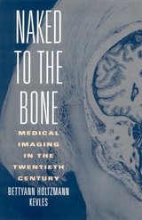
Naked to the Bone
Medical Imaging in the Twentieth CenturyKevles, Bettyann Holtzman
Rutgers University Press, 1996
X-rays, fluoroscopy, ultrasound, CT, MRI, and PET scans--medical imaging has become a familiar part of modern health care today. A century ago, however, the idea of looking inside the living body seemed absurd. Wilhelm Roentgen's X-ray image of his wife's shadowy hand--with her wedding band "floating" around a white bone--convinced doctors to rush the new tool into use for diagnosis and treatment.
By the 1920s, the technology was a commonplace wonder: army recruits had routinely lined up for chest X-rays during World War I, and children delighted in seeing the bones of their feet in the green glow of shoestore fluoroscopes. By the late 1960s, the computer and television were linked to produce medical images that were as startling as Roentgen's original X-rays. Computerized tomography (CT) and magnetic resonance imaging (MR) made it possible to picture soft tissues invisible to ordinary X-rays. Ultrasound allowed expectant parents to see their unborn children. Positron emission tomography (PET) enabled neuroscientists to map the brain.
In this lively history of medical imaging, the first to cover the full scope of the field from X-rays to MR-assistant surgery, Bettyann Kevles explores the consequences of these developments for medicine and society. Through lucid prose, vivid anecdotes, and more than seventy striking illustrations, she shows how medical imaging has transformed the practice of medicine--from pediatrics to dentistry, neurosurgery to geriatrics, gynecology to oncology.
Despite their formidable power to reveal the inner secrets of the body, no form of medical imaging can claim to be the product of a technological imperative. As Kevles points out, few of these costly inventions made it easily to the marketplace, and all are vulnerable to the changing economics of the health-care system. In the early years of X-rays, many doctors, technicians, and patients died from overexposure to the invisible radiation. Although we may still find delayed repercussions from these newer technologies, a different kind of danger may lie in our conviction that an early diagnosis is equivalent to a cure.
Beyond medicine, Kevles describes how X-rays and the newer technologies have become part of the texture of modern life and culture. They helped undermine Victorian sexual sensibilities, gave courts new forensic tools, provided plots for novels and movies, and offered artists from Picasso to Warhol new ways to depict the human form.
Naked to the Bone offers readers an unparalled picture of a key technology of the twentieth century.

Origins and Development of Medical Imaging
T. Doby and G. Alker
Southern Illinois University Press, 1997
For thousands of years, horror of the dead, superstition, and oppressive decrees prevented our ancestors from looking inside the human body; in ancient civilizations, diagnostics were based on imagination and theory, with only limited observation. So people developed suppositions about health and disease without knowing how the liver, heart, brain, and blood vessels looked or functioned. In tracing the history of medical imaging, Doby and Alker establish that it was not until the Renaissance and the detailed drawings of human anatomy by da Vinci and Vesalius that successful internal imaging of the human body was born.
At the turn of the nineteenth century, Roentgen’s discovery of the X-ray provided the first miraculous look into the living body. From that instant, medical imaging developed at an ever-increasing pace, evolving to more recent discoveries in nuclear medicine, CT scanning, and ultrasound and magnetic resonance imaging.
As Doby and Alker note, we can expect our efforts to be understood in the future only if we examine past events with an appreciation of the difficulties and challenges that faced our predecessors. Despite almost unbelievable advances in current medical technology, we must still rely on our own resources and common sense as human beings in understanding what technology can and cannot do for us.
A comprehensive set of appendixes pictorially depicting the history of imaging round out the volume.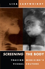
Screening The Body
Tracing Medicine’s Visual CultureLisa Cartwright
University of Minnesota Press, 1995
Moving images are used as diagnostic tools and locational devices every day in hospitals, clinics, and laboratories. But how and when did they come to be established and accepted sources of knowledge about the body in medical culture? How are the specialized techniques and codes of these imaging techniques determined, and whose bodies are studied, diagnosed, and treated with the help of optical recording devices?
Screening the Body traces the fascinating history of scientific film during the late nineteenth and early twentieth centuries to show that early experiments with cinema are important precedents of contemporary medical techniques such as ultrasound and PET scanning. Lisa Cartwright brings to light eccentric projects in the history of science and medicine, such as Thomas Edison's sensational attempt to image the brain with X rays before a public audience, and the efforts of doctors to use the motion picture camera to capture movements of the body, from the virtually imperceptible flow of blood to epileptic seizures.Drawing on feminist film theory, cultural studies, the history of film, and the writings of Foucault, Cartwright illustrates how this scientific cinema was part of a broader tendency in society toward the technological surveillance, management, and physical transformation of the individual body and the social body. She unveils an area of film culture that has rarely been discussed but that will leave readers with a new way of seeing the everyday practice of diagnostic imaging that we all inevitably encounter in clinics and hospitals.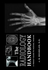
The Radiology Handbook
A Pocket Guide to Medical ImagingJ. S. Benseler
Ohio University Press, 2006
Designed for busy medical students, The Radiology Handbook is a quick and easy reference for any practitioner who needs information on ordering or interpreting images.
The book is divided into three parts:
- Part I presents a table, organized from head to toe, with recommended imaging tests for common clinical conditions.
- Part II is organized in a question and answer format that covers the following topics: how each major imaging modality works to create an image; what the basic precepts of image interpretation in each body system are; and where to find information and resources for continued learning.
- Part III is an imaging quiz beginning at the head and ending at the foot. Sixty images are provided to self-test knowledge about normal imaging anatomy and common imaging pathology.
Published in collaboration with the Ohio University College of Osteopathic Medicine, The Radiology Handbook is a convenient pocket-sized resource designed for medical students and non radiologists.