
 A SCHOLARLY BOOK REPOSITORY
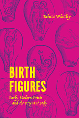
Birth figures are printed images of the pregnant womb, always shown in series, that depict the variety of ways in which a fetus can present for birth. Historian Rebecca Whiteley coined the term and here offers the first systematic analysis of the images’ creation, use, and impact. Whiteley reveals their origins in ancient medicine and explores their inclusion in many medieval gynecological manuscripts, focusing on their explosion in printed midwifery and surgical books in Western Europe from the mid-sixteenth to the mid-eighteenth century. During this period, birth figures formed a key part of the visual culture of medicine and midwifery and were widely produced. They reflected and shaped how the pregnant body was known and treated. And by providing crucial bodily knowledge to midwives and surgeons, birth figures were also deeply entangled with wider cultural preoccupations with generation and creativity, female power and agency, knowledge and its dissemination, and even the condition of the human in the universe.
Birth Figures studies how different kinds of people understood childbirth and engaged with midwifery manuals, from learned physicians to midwives to illiterate listeners. Rich and detailed, this vital history reveals the importance of birth figures in how midwifery was practiced and in how people, both medical professionals and lay readers, envisioned and understood the mysterious state of pregnancy.
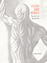
For centuries, anatomy was a fundamental component of artistic training, as artists such as Leonardo da Vinci and Michelangelo sought to skillfully portray the human form. In Europe, illustrations that captured the complex structure of the body—spectacularly realized by anatomists, artists, and printmakers in early atlases such as Andreas Vesalius’s De humani corporis fabrica libri septem of 1543—found an audience with both medical practitioners and artists.
Flesh and Bones examines the inventive ways anatomy has been presented from the sixteenth through the twenty-first century, including an animated corpse displaying its own body for study, anatomized antique sculpture, spectacular life-size prints, delicate paper flaps, and 3-D stereoscopic photographs. Drawn primarily from the vast holdings of the Getty Research Institute, the over 150 striking images, which range in media from woodcut to neon, reveal the uncanny beauty of the human body under the skin.
This volume is published to accompany an exhibition on view at the Getty Research Institute at the Getty Center from February 22 to July 10, 2022.
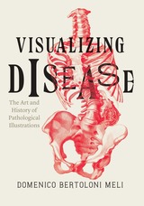
Starting in the Renaissance, Bertoloni Meli delves into the wide range of figures involved in the early study and representation of disease, including not just men of medicine, like anatomists, physicians, surgeons, and pathologists, but also draftsmen and engravers. Pathological preparations proved difficult to preserve and represent, and as Bertoloni Meli takes us through a number of different cases from the Renaissance to the mid-nineteenth century, we gain a new understanding of how knowledge of disease, interactions among medical men and artists, and changes in the technologies of preservation and representation of specimens interacted to slowly bring illustration into the medical world.
READERS
Browse our collection.
PUBLISHERS
See BiblioVault's publisher services.
STUDENT SERVICES
Files for college accessibility offices.
RECENTLY PUBLISHED


UChicago Accessibility Resources
home | accessibility | search | about | contact us
BiblioVault ® 2001 - 2025
The University of Chicago Press