front cover of Clinical Trials in Ovarian Cancer
Clinical Trials in Ovarian Cancer
Walsh, Christine S
Rutgers University Press, 2017
When a patient is diagnosed with a gynecological malignancy, she and her doctors must make urgent, high-risk decisions about her course of treatment. In selecting an appropriate plan of care, physicians must weigh the patient’s individual needs, the tumor’s specific characteristics, and the treatment’s potential side effects. Because there is no one-size-fits-all treatment solution, a plethora of clinical trials have been performed on ovarian cancer patients, but clinicians may struggle to keep up with this ever-growing body of research.   
 
Collecting and synthesizing research findings from a wide array of medical journal articles and book chapters, Clinical Trials in Ovarian Cancer provides physicians with an invaluable resource. Gynecologic oncologist Christine S. Walsh systematically outlines each of the seminal Phase III trials that have shaped the treatment of ovarian cancers, detailing the rationale for the trial, the patient population studied, treatment delivery methods, efficacy, toxicity, and trial conclusions. She provides a clear overview of established treatments, as well as still-controversial experimental approaches. 
 
The first book to organize this cutting-edge research into an easy-to-use reference, Clinical Trials in Ovarian Cancer should help medical personnel at all levels provide their patients with the highest standard of care. 
 
[more]

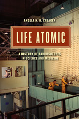
front cover of Life Atomic
Life Atomic
A History of Radioisotopes in Science and Medicine
Angela N. H. Creager
University of Chicago Press, 2013
After World War II, the US Atomic Energy Commission (AEC) began mass-producing radioisotopes, sending out nearly 64,000 shipments of radioactive materials to scientists and physicians by 1955. Even as the atomic bomb became the focus of Cold War anxiety, radioisotopes represented the government’s efforts to harness the power of the atom for peace—advancing medicine, domestic energy, and foreign relations.
           
In Life Atomic, Angela N. H. Creager tells the story of how these radioisotopes, which were simultaneously scientific tools and political icons, transformed biomedicine and ecology. Government-produced radioisotopes provided physicians with new tools for diagnosis and therapy, specifically cancer therapy, and enabled biologists to trace molecular transformations. Yet the government’s attempt to present radioisotopes as marvelous dividends of the atomic age was undercut in the 1950s by the fallout debates, as scientists and citizens recognized the hazards of low-level radiation. Creager reveals that growing consciousness of the danger of radioactivity did not reduce the demand for radioisotopes at hospitals and laboratories, but it did change their popular representation from a therapeutic agent to an environmental poison. She then demonstrates how, by the late twentieth century, public fear of radioactivity overshadowed any appreciation of the positive consequences of the AEC’s provision of radioisotopes for research and medicine.
[more]

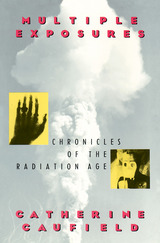
front cover of Multiple Exposures
Multiple Exposures
Chronicles of the Radiation Age
Catherine Caufield
University of Chicago Press, 1990
"Catherine Caufield has written an important book on an important topic:
the history behind the safety standards limiting the effects of high energy
radiation on human beings. . . . Provides an immense amount of information
in a very readable form."—W. Alan Runciman, Prometheus

"From fallout and radon to radioactive smoke detectors and dental X-rays,
Caufield traces the proliferation of the uses of radiation in medicine,
industry and the military, and in generating energy. An intelligent,
non-alarmist history."—Publishers Weekly
[more]

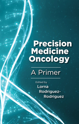
front cover of Precision Medicine Oncology
Precision Medicine Oncology
A Primer
Lorna Rodriguez-Rodriguez
Rutgers University Press, 2019
Precision medicine is rapidly becoming the standard-of-care for the treatment of cancer patients. This is made possible, in part, by the ready availability and reasonable costs of comprehensive DNA and RNA sequencing assays. However, precision medicine is complex and incorporates entirely new types of data and treatment paradigms that are outside of the training of most oncologists in practice today.

Precision Medicine Oncology: A Primer is a concise review of the fundamental principles and applications of precision medicine, intended for clinicians, particularly those working in oncology. It provides an accessible introduction to the technological advances in DNA and RNA sequencing, gives a detailed overview of approaches to the interpretation of molecular test results and their point-of-care implementation for individual patients, and describes innovative clinical trial designs in oncology as well as characteristics of the computational infrastructures through which massive quantities of data are collected, stored, and used in precision medicine oncology.
 
[more]

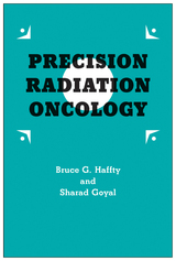
front cover of Precision Radiation Oncology
Precision Radiation Oncology
Haffty, Bruce G
Rutgers University Press, 2018
Precision medicine is a rapidly-evolving field in the management of cancer. The use of novel molecular or genetic signatures in local-regional management is still in its infancy. Precision Radiation Oncology demystifies this state-of-the-art research and technology. 

By describing current existing clinical and pathologic features, and focusing on the ability to improve outcomes in cancer using radiation therapy, this book discusses incorporating novel genomic- or biology-based biomarkers in the treatment of patients moving radiation oncology into precision/personalized medicine. Precision Radiation Oncology provides readers with an overview of the new developments of precision medicine in radiation oncology, further advancing the integration of new research findings into individualized radiation therapy and its clinical applications.  
 
[more]

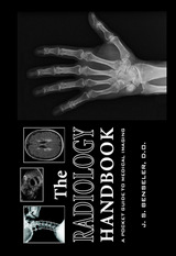
front cover of The Radiology Handbook
The Radiology Handbook
A Pocket Guide to Medical Imaging
J. S. Benseler
Ohio University Press, 2006

Designed for busy medical students, The Radiology Handbook is a quick and easy reference for any practitioner who needs information on ordering or interpreting images.

The book is divided into three parts:

- Part I presents a table, organized from head to toe, with recommended imaging tests for common clinical conditions.

- Part II is organized in a question and answer format that covers the following topics: how each major imaging modality works to create an image; what the basic precepts of image interpretation in each body system are; and where to find information and resources for continued learning.

- Part III is an imaging quiz beginning at the head and ending at the foot. Sixty images are provided to self-test knowledge about normal imaging anatomy and common imaging pathology.

Published in collaboration with the Ohio University College of Osteopathic Medicine, The Radiology Handbook is a convenient pocket-sized resource designed for medical students and non radiologists.
[more]

logo for Harvard University Press
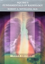
Squire’s Fundamentals of Radiology
Fifth Edition
Robert A. Novelline, M.D.
Harvard University Press, 1997

This new edition of Fundamentals of Radiology brings up to date the consummate, classic work by Lucy Frank Squire and Robert A. Novelline that has introduced generations of medical students to radiology.

The standard introductory text for more than thirty years, Fundamentals of Radiology is a model of clarity and comprehensiveness. Robert Novelline continues that tradition by thoroughly updating and expanding this edition to reflect the latest types and uses of imaging techniques. Complementing the text are many superb reproductions of plain film, computed tomography, magnetic-resonance, and ultrasound images--hundreds of them new to this edition. In addition, Novelline has added five important chapters. A new chapter near the beginning of the book provides an atlas of drawings and images that allows the reader to review normal plain film and CT anatomy. Another new chapter is devoted to vascular imaging, including CT angiography, MR angiography, and vascular ultrasound, especially full-color Doppler ultrasound images. The chapter on interventional radiology covers therapeutic procedures performed by radiologists, such as angioplasty, embolization, and percutaneous biopsy. To address the different medical conditions of males, females, and children, another new chapter examines the imaging of obstetrical, gynecological, testicular, prostate, and urethral disorders, and considers a variety of childhood ailments and problems, including child abuse. Finally, a new chapter on TB and AIDS shows how radiology can track the course of a single disease over time and trace the depredations of a multisystem disease.
[more]

logo for Harvard University Press
Squire’s Fundamentals of Radiology
Seventh Edition
Robert A. Novelline, M.D.
Harvard University Press, 2018

Medical students preparing for a career in clinical practice must become familiar with a wide range of diagnostic imaging techniques and image-guided interventions. They must learn to identify the indications for radiological examination and recognize the role each procedure plays in the workup, diagnosis, and therapeutic management of patients. That is why Squire’s Fundamentals of Radiology has been such an important, long-standing resource for medical students, physicians, and other professionals at all stages of their careers. It teaches essential topics in the radiology curriculum and features hundreds of illustrative cases clinicians can turn to again and again in practice.

In this long-awaited seventh edition, Robert Novelline provides more than 600 new high-resolution images representing the current breadth of radiological procedures: conventional x-rays, ultrasound, computed tomography (CT), magnetic resonance imaging (MRI), angiography, radioisotope scanning, positron emission tomography (PET), and molecular imaging. This edition’s expanded coverage addresses dual energy CT, breast tomosynthesis, PET-MR scanning, and tractography brain imaging, along with best practices for managing patient experiences during and after examination. All new images were produced at a major teaching hospital using state-of-the-art imaging technologies.

Squire’s Fundamentals of Radiology is designed to be read cover to cover by students, with concepts, principles, and methods progressing in a logical, cumulative manner. It also serves as an invaluable tool for teachers and an indispensable reference for seasoned practitioners. Written by a radiologist who has trained thousands of medical students and residents, this textbook is the clear choice for excelling in the general practice of radiology.

[more]

logo for Harvard University Press
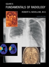
Squire’s Fundamentals of Radiology
Sixth Edition
Robert A. Novelline, M.D.
Harvard University Press
In the past five years, the development of new imaging technologies that make possible faster and more accurate diagnoses has significantly improved the imaging of disease and injury. This new edition of Squire’s Fundamentals of Radiology describes and illustrates these new techniques to prepare medical students and other radiology learners to provide the most optimal and up-to-date imaging management for their patients. Not only are new diagnostic techniques outlined, such as the multidetector computed tomography diagnosis of pulmonary embolism and the diffusion-weighted magnetic-resonance imaging of stroke, but hundreds of new diagnostic images have been included to illustrate the radiological characteristics of common diseases with state-of-the-art computed radiography, ultrasound, multidetector computed tomography, and magnetic-resonance images. The text has been completely reviewed and updated to present the latest and best strategies in diagnostic imaging.New interventional radiology procedures have been added, including vertebroplasty, a percutaneous injection treatment of painful spinal compression fractures; uterine artery embolization, a surgical alternative to hysterectomy in women with painful or bleeding uterine fibroids; and radiofrequency ablation, a percutaneous technique for treating unresectable tumors in the liver and other organs with probes that superheat and thus destroy cancer cells.A new chapter on advances in diagnostic imaging describes many cutting-edge imaging technologies, such as three-dimensional and digital imaging, functional magnetic-resonance imaging, PET–CT (positron emission tomography combined with computed tomography), cardiac calcium CT scoring, multidetector gated cardiac CT, and molecular imaging.
[more]


Send via email Share on Facebook Share on Twitter